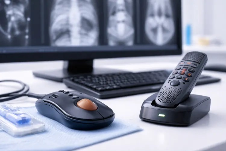
Radiology stands at the forefront of the healthcare data revolution. With billions of medical images (X-rays, CT scans, MRIs, Ultrasounds) generated globally each year, modern radiology departments face a critical bottleneck: a massive increase in imaging volume coupled with an ongoing shortage of trained specialists.
This workload pressure leads to delayed reports, potential burnout, and the risk of missing subtle, critical findings.
Artificial Intelligence (AI), specifically through deep learning and computer vision, is now moving beyond research and into the Picture Archiving and Communication System (PACS), serving as an indispensable AI co-pilot for the modern radiologist. AI tools automate time-consuming tasks, prioritize life-threatening findings, and provide quantitative analysis that enhances diagnostic confidence.
Atlas Meditech’s AtlasGPT is an intelligent system designed to streamline the entire diagnostic imaging process. Integrating across various modalities, AtlasGPT provides real-time decision support, helping radiologists manage high volumes while focusing their attention on the most complex cases.
Key Applications of AI in Diagnostic Radiology
AI algorithms are transforming radiology across three major domains: workflow efficiency, diagnostic accuracy, and quantitative analysis.
1. Workflow Triage and Prioritization
Perhaps the most immediate benefit of AI is its ability to act as a virtual gatekeeper, managing the queue of incoming scans to ensure life-threatening conditions are addressed immediately.
Critical Finding Alerts: AI instantly analyzes scans (e.g., Head CTs, Chest X-rays) for acute findings such as intracranial hemorrhage, pulmonary embolism (PE), or aortic dissection.
Smart Worklist Prioritization: The AI system dynamically reorders the radiologist's worklist, pushing studies with potential critical findings to the top. This enhances overall departmental efficiency and ensures patient safety in busy environments.
2. Enhanced Detection and Diagnostic Accuracy
AI algorithms are trained on vast datasets of annotated images, allowing them to identify patterns that might be too subtle or consistent to be easily noticed by the human eye.
Cancer Screening: In mammography, AI-powered tools assist in detecting malignant lesions and assessing breast density, significantly lowering both false positives and false negatives, especially in cases of dense breast tissue.
Subtle Fracture Detection: In trauma cases, AI has proven highly effective at identifying occult (hidden) fractures in musculoskeletal imaging that are often missed on initial review.
Neuroimaging: AI is crucial in rapidly detecting and segmenting brain tumors, assessing neurodegenerative diseases, and quantifying disease progression, often achieving high accuracy in classifying tumor grades from MRI data.
3. Quantitative Analysis and Reporting
AI introduces objectivity into the interpretation process through Radiomics and automation.
Automated Segmentation: AI automates the laborious task of segmenting (outlining) organs and lesions, which is critical for measuring tumor volume, assessing treatment response, and planning radiation therapy. This cuts preparation time dramatically.
Quantitative Reporting: AI moves reporting from purely descriptive to quantitative. It generates objective measurements of lesions, tissue characteristics, and functional metrics, which leads to more standardized, consistent reports across a practice.
Generative AI in Reporting: Large Language Models (LLMs) and Generative AI can draft initial radiology reports based on image findings and dictated notes, streamlining documentation and allowing radiologists to focus on complex interpretation and consultation.
The AtlasGPT Advantage in Radiology
AtlasGPT is built to be the comprehensive, multi-modal AI solution for radiologists:
Feature | Description |
Real-time Cross-Modality Analysis | Integrates and analyzes data from X-ray, CT, MRI, and patient records to provide a holistic view and flag potential discrepancies. |
Intelligent Alerting | Implements FDA-cleared and proprietary algorithms to prioritize acute pathologies (e.g., PE, stroke, pneumothorax) and ensure rapid clinical action. |
Standardized Quantification | Provides automated, objective measurements for tumor size, nodule density, and anatomical segmentation, reducing inter-reader variability. |
Generative Report Drafting | Leverages advanced LLMs to draft structured, quantitative reports and automatically pull relevant historical data from the EHR. |
FAQs About Radiology AI Tools
What is Radiomics?
Radiomics uses AI to extract a vast amount of quantitative data (features) from medical images that are not visible to the naked eye. This data can be used to predict tumor response, prognosis, and patient outcomes.
Does AI replace the radiologist?
No. AI is designed to augment the radiologist's ability, handling routine and high-volume tasks. The radiologist is still essential for complex interpretation, patient context integration, and final clinical judgment.
How does AI improve MRI scan times?
AI-based Deep Learning (DL) image reconstruction methods can accelerate the acquisition process by filling in data gaps, reducing the total time a patient spends in the scanner while maintaining or improving image quality.
What is the main challenge of adopting AI in radiology?
Key challenges include ensuring data quality and diversity for training models, addressing data privacy concerns, and securing seamless, compliant integration with existing PACS and EHR systems.
Transform Your Imaging Workflow with AtlasGPT
The synergy between the radiologist's expertise and AI's computational power is the future of medical imaging. By adopting solutions like AtlasGPT, institutions can significantly reduce turnaround times, increase diagnostic precision, and ensure that every patient receives the fastest, most accurate diagnosis possible.
Experience AtlasGPT in Radiology today and lead the way in AI-augmented diagnostic care.